Präsentation herunterladen
Die Präsentation wird geladen. Bitte warten

1
Wie entstehen Knochenmetastasen?
Martin Pecherstorfer, Landesklinikum Krems

2
Der Knochen ähnelt einem Stahlgerüst!

3
Aufbau und Abbau – eine Großbaustelle!

4
Einmal zerstörter Knochen kann nicht vollständig wiederhergestellt werden

5
Osteoklasten bauen den Knochen ab
Osteoclast Key Point This slide shows a scanning electron micrograph of an osteoclast-resorbing bone.1 Supplementary Information Osteoclasts differentiate from haematopoietic stem cells. Their function is bone resorption and so they play a vital role in skeletal development, growth, maintenance and calcium metabolism.2 They resorb both the mineral and organic phases of bone by attaching to the bone matrix, polarising, and forming an irregular border at the bone surface to isolate the resorption compartment.2 Bone mineral dissolution then proceeds through secretion of acids and collagen-degrading enzymes onto the bone surface.2 Once the resorption products have been degraded, the osteoclast either relocates to another site (as depicted in the electron micrograph above), or undergoes apoptosis.2 The collagen fragments, and solubilised calcium and phosphate generated by bone degradation, are released into the circulation.3 Research has identified RANK Ligand as essential mediator of osteoclast formation, function and survival. 1. Eelectron micrograph reproduced with the permission of The Bone Research Society; Accessed 2/19/2009. 2. Bar-Shavit Z. J Cell Biochem 2007;102: 3. Boyle WJ, et al. Nature 2003;423: Bone surface resorbed by osteoclast Adapted from: Electron micrograph photo reproduced with permission.© Tim Arnett, The Bone Research Society.
, or undergoes apoptosis.2. The collagen fragments, and solubilised calcium and phosphate generated by bone degradation, are released into the circulation.3. Research has identified RANK Ligand as essential mediator of osteoclast formation, function and survival. 1. Eelectron micrograph reproduced with the permission of The Bone Research Society; Accessed 2/19/ Bar-Shavit Z. J Cell Biochem 2007;102: Boyle WJ, et al. Nature 2003;423: Bone surface resorbed by osteoclast. Adapted from: Electron micrograph photo reproduced with permission.© Tim Arnett, The Bone Research Society.")
6
“Lytisches” Knochen-Microenvironment: Erhöhte Osteoklastenaktivität
RANKL RANK RANKL Osteoblasten Osteoklasten

7
Pre-Fusion Osteoclast
RANK Ligand ist ein essentieller Mediator der Osteoklastenformation, -funktion und Überleben Pre-Fusion Osteoclast CFU-M RANK Ligand RANK Multinucleated Osteoclast Hormones Growth factors Cytokines Activated Osteoclast Aktivierter Osteoklast Osteoblasts Bone Formation Adapted from: Boyle et al., Nature 2003 Bone Resorption Boyle WJ et al. Nature 2003;423:337–342

8
Pre-Fusion Osteoclast
Osteoprotegerin (OPG) verhindert RANKL Bindung an RANK und hemmt Osteoklastenformation, Function und Überleben Pre-Fusion Osteoclast CFU-M RANK Ligand RANK Osteoprotegerin Hormones Growth factors Cytokines Osteoclast Formation, Function, and Survival Inhibited Osteoblasts Knochenformation Knochenresorption gehemmt Boyle WJ et al. Nature 2003;423:337–342
 verhindert RANKL Bindung an RANK und hemmt Osteoklastenformation, Function und Überleben. Pre-Fusion Osteoclast. CFU-M. RANK Ligand. RANK. Osteoprotegerin. Hormones Growth factors. Cytokines. Osteoclast Formation, Function, and Survival Inhibited. Osteoblasts. Knochenformation. Knochenresorption gehemmt. Boyle WJ et al. Nature 2003;423:337–342.")
9
Verminderte Knochendichte (BMD) Microarchitektur-störungen
Eine Imbalance in der Knochenresorption/Formation schwächt trabekulären Knochen Verminderte Knochendichte (BMD) Microarchitektur-störungen Erhöhte Frakturgefahr Normaler Knochen Osteoporotischer Knochen Photos reproduced from Dempster DW et al. J Bone Miner Res. 1986; 1: 15–21 with permission of the American Society for Bone and Mineral Research
 Microarchitektur-störungen. Erhöhte Frakturgefahr. Normaler Knochen. Osteoporotischer Knochen. Photos reproduced from Dempster DW et al. J Bone Miner Res. 1986; 1: 15–21. with permission of the American Society for Bone and Mineral Research.")
10
Knochen-Metastasen

11
Kommunikation über das periphere Blut
Manche Krebszellen (z.B. Brustkrebszellen) haben eine spezielle Beziehung zum Knochen! Knochemark Stammzellen Immuneffektorzellen Antigenpräsentierende Zellen Tumor Kommunikation über das periphere Blut
 haben eine spezielle Beziehung zum Knochen! Knochemark. Stammzellen. Immuneffektorzellen. Antigenpräsentierende Zellen. Tumor. Kommunikation über das periphere Blut.")
12
Der Zielort der Metastasierung hängt nicht vom Zufall sondern von der enzymatischen Ausstattung der Krebszelle ab

13
Der Teufelskreis bei osteolytischen Metastasen
Bisphosphonates Overview Der Teufelskreis bei osteolytischen Metastasen Roodman, G. D. N Engl J Med 2004;350: 13

14
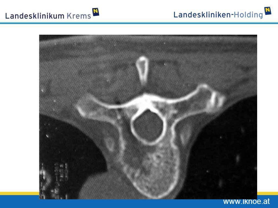
Radiologisch unterscheidet man:
Lytische Knochenherde (Myelom, Nierenzell-karzinom) Gemischte Knochenmetastasen (Mamma Ca) Osteoblastische Knochenherde (Prostata Ca)
 Gemischte Knochenmetastasen (Mamma Ca) Osteoblastische Knochenherde (Prostata Ca)")
15
Beim Prostatakrebs werden nicht nur die Osteoklasten sondern auch die Osteoblasten aktiviert

16
Osteoblastische und osteoklastische Metastasen der Schädeldecke
Cruveilhier ( )
")
17
Osteoblastische Metastasen
Soft X-ray SR-µCT Gesunde Wirbelkörper1 X-ray images of osteoblastic metastasis from prostate cancer appear radiodense, however synchrotron radiation (SR)-µCT analysis of the microarchitecture and mineralization of osteoblastic metastasis reveal the disorganized woven bone and the deterioration of the trabecular structure that provides strength in the normal bone.8 Representative images are shown in the slide: The top panel shows a normal control subject. The soft X ray and 3D volume-rendered SR-μCT images of normal trabecular bone from the L2 vertebral body taken from a 37-year-old man. The organized trabecular structure can be observed in the soft x-ray. The 3D image clearly shows both rod- and plate-like structures of the trabecular bone. The bottom panel shows a patient with osteoblastic metastasis from prostate cancer. The soft X ray and 3D volume-rendered SR-μCT images of metastatic trabecular bone from the L3 vertebral body, taken from a 67-year-old man with bone metastases from prostate cancer. The irregular, disorganized woven bone structure can be observed in the osteoblastic bone lesion. The 3D image shows an increased number of trabeculae with an irregular surface. The trabecular number was greater than that in normal tissue, whereas no difference in trabecular thickness was observed. Although the osteoblastic lesion in the soft x-ray on this slide appears radiographically opaque due to the increase in bone volume1, the increased bone volume is a result of formation of woven bone. This woven bone is poorly mineralized with insufficient mechanical strength1-5 due to increased bone turnover as compared to the normal bone6,7. Clarke NW, et al. Br J Urol. 1991;68:74-80. Charhon SA, et al. Cancer. 1983;51: Roudier M, et al. J Clin Invest In Review. Roodman GD, Windle JJ. J Clin Invest. 2005;115: Martin RB, et al. Skeletal Biology. In: Skeletal Tissue Mechanics. 1989;29-78. Kanis JA, McCloskey EV. Cancer. 1997;80:1538–1545. Urwin GH, et al. Br J Urol. 1985;57: Sone T, et al. Bone. 2004;35: Soft X-ray SR-µCT Osteoblastische Metastasen
-µCT analysis of the microarchitecture and mineralization of osteoblastic metastasis reveal the disorganized woven bone and the deterioration of the trabecular structure that provides strength in the normal bone.8. Representative images are shown in the slide: The top panel shows a normal control subject. The soft X ray and 3D volume-rendered SR-μCT images of normal trabecular bone from the L2 vertebral body taken from a 37-year-old man. The organized trabecular structure can be observed in the soft x-ray. The 3D image clearly shows both rod- and plate-like structures of the trabecular bone. The bottom panel shows a patient with osteoblastic metastasis from prostate cancer. The soft X ray and 3D volume-rendered SR-μCT images of metastatic trabecular bone from the L3 vertebral body, taken from a 67-year-old man with bone metastases from prostate cancer. The irregular, disorganized woven bone structure can be observed in the osteoblastic bone lesion. The 3D image shows an increased number of trabeculae with an irregular surface. The trabecular number was greater than that in normal tissue, whereas no difference in trabecular thickness was observed. Although the osteoblastic lesion in the soft x-ray on this slide appears radiographically opaque due to the increase in bone volume1, the increased bone volume is a result of formation of woven bone. This woven bone is poorly mineralized with insufficient mechanical strength1-5 due to increased bone turnover as compared to the normal bone6,7. Clarke NW, et al. Br J Urol. 1991;68: Charhon SA, et al. Cancer. 1983;51: Roudier M, et al. J Clin Invest In Review. Roodman GD, Windle JJ. J Clin Invest. 2005;115: Martin RB, et al. Skeletal Biology. In: Skeletal Tissue Mechanics. 1989; Kanis JA, McCloskey EV. Cancer. 1997;80:1538–1545. Urwin GH, et al. Br J Urol. 1985;57: Sone T, et al. Bone. 2004;35: Soft X-ray. SR-µCT. Osteoblastische Metastasen.")
18
Osteolytische Metastase des Oberarms

19
Osteoblastische Durchsetzung des Beckens bei Prostata Ca

20
Knochenschmerz ist das Leitsymptom bei Knochenmetastasen
Inzidenz von Knochenmetastasen Mammakarzinom 65–75% Prostatakarzinom 65–75% Multiples Myelom 70–95% Bronchialkarzinom 30–40%1 Typische Komplikationen Knochenschmerz ~50–90% pathologische Frakturen 10–40% Hyperkalzämie <10%2

21
Wie entseht der Schmerz bei Knochenmetastase?
Entzündungszellen Knochen Dorsales Spinalganglion Zum Gehirn IL-1b TNF-a NGF PGE2 Sensor. Neuron + Osteoklasten + + + + + + + + NGF + PGE2 Tumorzellen ET-1 Rückenmark

22
Dichtes Geflecht von sensiblen Nerven in Knochenmark und Periost

23
Knochenschmerzen beeinflussen Lebensqualtät und Morbidität
Arztbesuche/ Therapieverfahren Eingeschränkte Lebensqualität Knochen- schmerz Nebenwirkungen der Therapie Abhängigkeit von Pflegepersonal Krankenhaus- Aufenthalte Immobilität (Bettlägerigkeit)
")
24
Behandlungsmöglichkeiten bei Knochenmetastasen
Zerstörung der Tumorzellen: Chemotherapie Strahlentherapie Antikörpertherapie Zerstörung der Osteoklasten: Bisphosphonate Denosumab

25
Denosumab ein, Antiköper gegen RANK Ligand
CFU-GM Pre-fusion osteoclast Osteoclast formation inhibited RANK OPG Denosumab Osteoclast function and survival inhibited Hormones Growth factors Cytokines Key Point The discovery of the RANK/RANK ligand/OPG pathway and its implications for the pathogenesis of osteoporosis provides a molecular target for new therapies to improve bone health. Inhibition of RANK Ligand would be offer the advantage that it would mimic the endogenous pathway for inhibiting the formation, function and survival of osteoclasts. Supplementary Information RANK Ligand is an essential mediator in the formation, activation, and survival of osteoclasts.1 OPG sequesters free RANK Ligand thus preventing its binding to the RANK receptor.1 A suitable therapy for osteoporosis would: Mimic the activity and binding specificity of OPG for RANK Ligand. Be recognised by the body as ‘self’. Not be regulated by the normal bone homeostasis pathways. 1. Boyle WJ, et al. Nature 2003;423: Osteoblasts Bone resorption inhibited

26
Effekte der Bisphosphonate und von Denosumab bei Knochenmetastasen
Schmerzreduktion Verminderte Häufigkeit pathologischer Frakturen Verminderte Häufigkeit hyperkalzämischer Episoden Verminderte Notwendigkeit der Strahlentherapie Verlängerung des Überlebens?

27
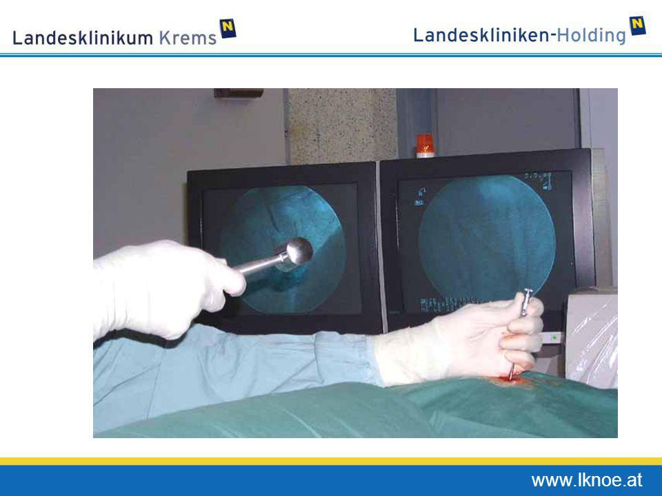
Vertebroplastik und Kyphoplastik

29
Nadel Pistole Verbindungsschlauch Aspirationskanüle

33
Vorgehen bei Kyphoplastik
Cannulation of the vertebral body Placement of the inflatable bone tamps (IBT) Inflation of IBT, restoration of vertebral body Resultant cavity and cement fill Dudeney S. et al., J Clin Oncol 2002
 Inflation of IBT, restoration of. vertebral body. Resultant cavity and cement fill. Dudeney S. et al., J Clin Oncol")
34
Vielleicht wird es uns einmal gelingen diese Probleme zu verhindern!
Beckenübersicht mit Osteolyse im Os pubis Humerus Femur Wirbelsäule mit pathol. Fraktur bei Osteolyse mit Osteolyse Quelle: Atlas der Radiologie,1997

38
Die Hypothese des “Teufelskreises” der Kommunikation Tumor - Knochen
RANKL RANK OPG Tumour cell PTHrP, BMP, TGF-β, IGF, FGF, VEGF, ET1, WNT PDGF, BMPs TGF-β, IGFs FGFs CA+2 The “vicious cycle” hypothesizes that tumor cells interact with the bone marrow microenvironment to drive bone destruction and tumor growth in a symbiotic relationship. Tumor cells secrete parathyroid-hormone-related peptide (PTHrP), which is the primary stimulator of osteoblast production of RANK Ligand1 PTHrP induces both the production of RANK Ligand and down-regulates OPG production by osteoblasts, thereby stimulating osteoclastogenesis2 Other factors produced and secreted by tumor cells (endothelin-1(ET-1), IL-6, prostaglandin E2, TNF, and macrophage colony-stimulating factor) also increase the expression of RANK Ligand1,4 The increased expression of RANK Ligand in the tumor environment leads to increased formation, activation and survival of osteoclasts, and resulting osteolytic lesions3 Osteolysis (process of bone resorption) then leads to the release of growth factors derived from bone, including: 1,2 transforming growth factor-β (TGF-β) insulin-like growth factors (IGFs) fibroblast growth factors (FGFs) platelet-derived growth factor (PDGF) bone morphogenetic proteins (BMPs) These factors increase the production of PTHrP or promote tumor growth directly1 Specifically, the growth factors bind to receptors on the surface of the tumor cells activating autophosphorylation and signaling through pathways involving SMAD (cytoplasmic mediators of most TGF-β signals) and mitogen-activated protein kinase (MAPK)2 Bone destruction increases local extracellular calcium (Ca2+) concentrations, which have also been shown to promote tumor growth and the production of PTHrP2 Thus, tumor-cell proliferation and production of PTHrP through the signaling of these pathways is promoted and the cycle continues Roodman GD, N Engl J Med ;350: Mundy GR, et al. Nature Reviews Cancer. 2002;2: Kitazawa S, et al. J Pathol. 2002;198:228–36. Guise TA, et al. Endocrin Rev. 1998;19:18-54. Activated osteoclast Osteoblasts adapted from Roodman D, N Engl J Med 2004; 350:1655.
, which is the primary stimulator of osteoblast production of RANK Ligand1. PTHrP induces both the production of RANK Ligand and down-regulates OPG production by osteoblasts, thereby stimulating osteoclastogenesis2. Other factors produced and secreted by tumor cells (endothelin-1(ET-1), IL-6, prostaglandin E2, TNF, and macrophage colony-stimulating factor) also increase the expression of RANK Ligand1,4. The increased expression of RANK Ligand in the tumor environment leads to increased formation, activation and survival of osteoclasts, and resulting osteolytic lesions3. Osteolysis (process of bone resorption) then leads to the release of growth factors derived from bone, including: 1,2. transforming growth factor-β (TGF-β) insulin-like growth factors (IGFs) fibroblast growth factors (FGFs) platelet-derived growth factor (PDGF) bone morphogenetic proteins (BMPs) These factors increase the production of PTHrP or promote tumor growth directly1. Specifically, the growth factors bind to receptors on the surface of the tumor cells activating autophosphorylation and signaling through pathways involving SMAD (cytoplasmic mediators of most TGF-β signals) and mitogen-activated protein kinase (MAPK)2. Bone destruction increases local extracellular calcium (Ca2+) concentrations, which have also been shown to promote tumor growth and the production of PTHrP2. Thus, tumor-cell proliferation and production of PTHrP through the signaling of these pathways is promoted and the cycle continues. Roodman GD, N Engl J Med. 2004;350: Mundy GR, et al. Nature Reviews Cancer. 2002;2: Kitazawa S, et al. J Pathol. 2002;198:228–36. Guise TA, et al. Endocrin Rev. 1998;19: Activated osteoclast. Osteoblasts. adapted from Roodman D, N Engl J Med 2004; 350:1655.")
39
RANKL Inhibition blockiert eine tumor-induzierte Osteolyse
4 MDA-231 intracardiac mouse model Radiographische Läsionen # lesions / mouse 2 * * 0.3 1 3 OPG dose (mg/kg) 0.3 1 3 60 120 180 OPG dose (mg/kg) * # of OC / mm2 tumour area In animal models with intracardiac injection of human MDA-231 breast cancer cells, the ability of OPG to inhibit tumor-induced osteoclastogenesis, and osteolysis was evaluated.1 The injection of MDA-231 breast cancer cells into the left ventricle of nude mice resulted in experimental bone metastases that were associated with osteolytic lesions. Mice were treated with OPG (at the indicated doses, 3 times per week for 4 weeks) starting immediately after tumor cell inoculation. RANK Ligand inhibition with OPG dose-dependently decreased the number and area of radiographically evident lytic bone lesions.1 OPG treatment at the highest doses completely prevented the radiographic appearance of osteolytic lesions (right radiograph). In the animals treated with OPG, note the increased radio-opacity of the tibial and femoral metaphyseal growth plate region associated with OPG treatment. This increase is a normal pharmacological response to OPG treatment by growing mice, whereby resorption of the secondary spongiosa is inhibited, and trabecular bone accumulates. RANK Ligand inhibition was also observed to significantly decrease the number of osteoclasts within the tumor, with a maximal reduction of 90% at 3 mg/kg (P < 0.01). In this model of experimental bone metastases, RANK Ligand inhibition was associated with significant reductions in both the frequency and the size of skeletal tumor nests and in preventing tumor-associated osteolysis.1 Morony S, et al. Osteoprotegerin Inhibits Osteolysis and Decreases Skeletal Tumor Burden in Syngeneic and Nude Mouse Models of Experimental Bone Metastasis. Cancer Res ;61: Control RANK ligand inhibition *Significantly different from 0 mg/kg OPG reproduced from Morony S, et al. Cancer Res 2001;61(11): with permission of the American Association for Cancer Research.
 OPG dose (mg/kg) * # of OC / mm2 tumour area. In animal models with intracardiac injection of human MDA-231 breast cancer cells, the ability of OPG to inhibit tumor-induced osteoclastogenesis, and osteolysis was evaluated.1 The injection of MDA-231 breast cancer cells into the left ventricle of nude mice resulted in experimental bone metastases that were associated with osteolytic lesions. Mice were treated with OPG (at the indicated doses, 3 times per week for 4 weeks) starting immediately after tumor cell inoculation. RANK Ligand inhibition with OPG dose-dependently decreased the number and area of radiographically evident lytic bone lesions.1 OPG treatment at the highest doses completely prevented the radiographic appearance of osteolytic lesions (right radiograph). In the animals treated with OPG, note the increased radio-opacity of the tibial and femoral metaphyseal growth plate region associated with OPG treatment. This increase is a normal pharmacological response to OPG treatment by growing mice, whereby resorption of the secondary spongiosa is inhibited, and trabecular bone accumulates. RANK Ligand inhibition was also observed to significantly decrease the number of osteoclasts within the tumor, with a maximal reduction of 90% at 3 mg/kg (P < 0.01). In this model of experimental bone metastases, RANK Ligand inhibition was associated with significant reductions in both the frequency and the size of skeletal tumor nests and in preventing tumor-associated osteolysis.1. Morony S, et al. Osteoprotegerin Inhibits Osteolysis and Decreases Skeletal Tumor Burden in Syngeneic and Nude Mouse Models of Experimental Bone Metastasis. Cancer Res. 2001;61: Control. RANK ligand inhibition. *Significantly different from 0 mg/kg OPG. reproduced from Morony S, et al. Cancer Res 2001;61(11): with permission of the American Association for Cancer Research.")
Ähnliche Präsentationen


>")


 bei Herzinsuffizienz>")


