Präsentation herunterladen
Die Präsentation wird geladen. Bitte warten

1
Maligne Lymphome R. Weide Praxisklinik für Hämatologie und Onkologie Koblenz

3
Maligne Lymphome Systematik der malignen Lymphome Grundlagen der Therapie Kasuistiken Zusammenfassung

4
Systematik: Lymphomentstehung
Pluripotente Stammzelle Myeloische Vorläuferzelle Lymphatische Vorläuferzelle Thymus Hochmaligne (aggressive) Lymphome Niedrigmaligne (indolente) Lymphome Chronisch lymphatische Leukämie B Lymphozyten T Lymphozyten
 Lymphome. Niedrigmaligne (indolente) Lymphome. Chronisch lymphatische Leukämie. B. Lymphozyten. T. Lymphozyten.")
5
Non-Hodgkin-Lymphome
Systematik: Histologie Morbus Hodgkin Non-Hodgkin-Lymphome Niedrigmaligne = Indolent Hochmaligne = Aggressiv

6
Systematik: Prognose Mediane Überlebenszeit von Indolente Lymphome:
einigen Jahren (~10) (B-CLL, follikuläre Lymphome Grad I + II) Aggressive Lymphome: Mediane Überlebenszeit von Monaten (Follikuläre Lymphome Grad III, diffus großzellige Lymphome, anaplastische Lymphome) Hochaggressive Lymphome: Mediane Überlebenszeit von Wochen (Burkitt Lymphom, B-ALL, T-ALL)
 (B-CLL, follikuläre. Lymphome Grad I + II) Aggressive Lymphome: Mediane Überlebenszeit von Monaten. (Follikuläre Lymphome Grad III, diffus großzellige Lymphome, anaplastische Lymphome) Hochaggressive Lymphome: Mediane Überlebenszeit von Wochen. (Burkitt Lymphom, B-ALL, T-ALL)")
7
Systematik: Stadieneinteilung: I - IV
KM I II III IV Das Stadium ist mitbestimmend für die Prognose! Niedriges Stadium hohe Heilungschancen Hohes Stadium niedrige Heilungschancen

8
Systematik: B-Symptome
A: Ohne B-Symptome B: Mit B-Symptomen B: Nachtschweiß Fieber Gewichtsverlust 10% in 6 Monaten

9
Systematik: Therapieziele
HEILUNG Morbus Hodgkin Hochmaligne NHL = Aggressive NHL HEILUNG Stadium I + II Niedrigmaligne NHL = Indolente NHL HEILUNG ? HEILUNG ? Stadium III + IV Palliativ

10
2. Grundlagen der Therapie
Strahlentherapie Chemotherapie Immunotherapie (Kalte Antikörper) Immunochemotherapie Radioimmunotherapie (Radioaktiv markierte Antikörper) Hochdosischemotherapie ± Ganzkörperbestrahlung mit anschließender autologer oder allogener Stammzelltransplantation
 Immunochemotherapie. Radioimmunotherapie (Radioaktiv markierte Antikörper) Hochdosischemotherapie ± Ganzkörperbestrahlung. mit anschließender autologer oder allogener Stammzelltransplantation.")
11
Grundlagen:Behandlungsmöglichkeiten
Strahlentherapie Chemotherapie (Zellgifte) Immunotherapie (Antikörper) CD20 Rituximab CD20 Rituximab Chemotherapie + Antikörper Y* CD20 Radioimmunotherapie
 Immunotherapie. (Antikörper) CD20. Rituximab. CD20. Rituximab. Chemotherapie. + Antikörper. Y* CD20. Radioimmunotherapie.")
12
Grundlagen der Therapie
Strahlentherapie Involved field Extended field Total nodal Ganzkörper (TBI)
")
13
Grundlagen der Therapie
Chemotherapie (Polychemotherapie) Alkylantien (z.B. Cyclophosphamid) Anthrazykline (z.B. Doxorubicin) Vinca - Alkaloide ( z.B. Vincristin) Purin - Antagonisten (z.B. Fludarabin) Steroide
 Alkylantien (z.B. Cyclophosphamid) Anthrazykline (z.B. Doxorubicin) Vinca - Alkaloide ( z.B. Vincristin) Purin - Antagonisten (z.B. Fludarabin) Steroide.")
14
Grundlagen der Therapie
Immunotherapie B-Zelle B + T-Zelle CD20 Rituximab c CD52 Campath c

15
Grundlagen der Therapie
Immunochemotherapie R-CHOP R = Rituximab C = Cyclophosphamid H = Hydroxydoxorubicin O = Oncovin = Vincristin P = Prednisolon

16
Grundlagen der Therapie
Radioimmunotherapie = Radioaktiv markierte Antikörper CD20 Zevalin B Y90

17
Grundlagen der Therapie
Hochdosischemotherapie ± Ganzkörperbestrahlung mit anschließender autologer Stammzelltransplantation

18
Grundlagen der Therapie
Hochdosischemotherapie ± Ganzkörperbestrahlung mit anschließender allogener Stammzelltransplantation

19
Hochmalignes (aggressives) Lymphom
 Lymphom")
20
Hochmaligne (aggressive) Lymphome müssen in jedem Stadium immer sofort mit einer Chemoimmunotherapie (Chemotherapie + Antikörper) behandelt werden.
 Lymphome müssen in jedem Stadium immer sofort mit einer Chemoimmunotherapie (Chemotherapie + Antikörper) behandelt werden.")
21
Hochmaligne (aggressive) Lymphome International Prognostic Index (IPI)
Alter > 60 Jahre Serum LDH erhöht ECOG Performance Status > 2 (Köperzustand) Stadium III oder IV Mehr als 1 Lokalisation eines Extranodalbefalls Für jedes Charakteristikum wird 1 Punkt vergeben: Punkte Risikogruppe Jahresüberleben Niedrig % Niedrig - intermediär % Hoch – intermediär % Hoch %
 Stadium III oder IV. Mehr als 1 Lokalisation eines Extranodalbefalls. Für jedes Charakteristikum wird 1 Punkt vergeben: Punkte Risikogruppe 5-Jahresüberleben. 0-1 Niedrig 73 % 2 Niedrig - intermediär 51 % 3 Hoch – intermediär 43 % 4-5 Hoch 26 %")
22
Maligne Lymphome - Heilungschancen
73% Hochmaligne (aggressive) IPI 0-1 51% IPI 2 43% IPI 3 26% IPI 4-5
 IPI % IPI 2. 43% IPI 3. 26% IPI 4-5.")
23
Wichtigste Grundlage für die Behandlung ist die feingewebliche Diagnosesicherung:
Bei Diagnose CT-gesteuerte Biopsie

24
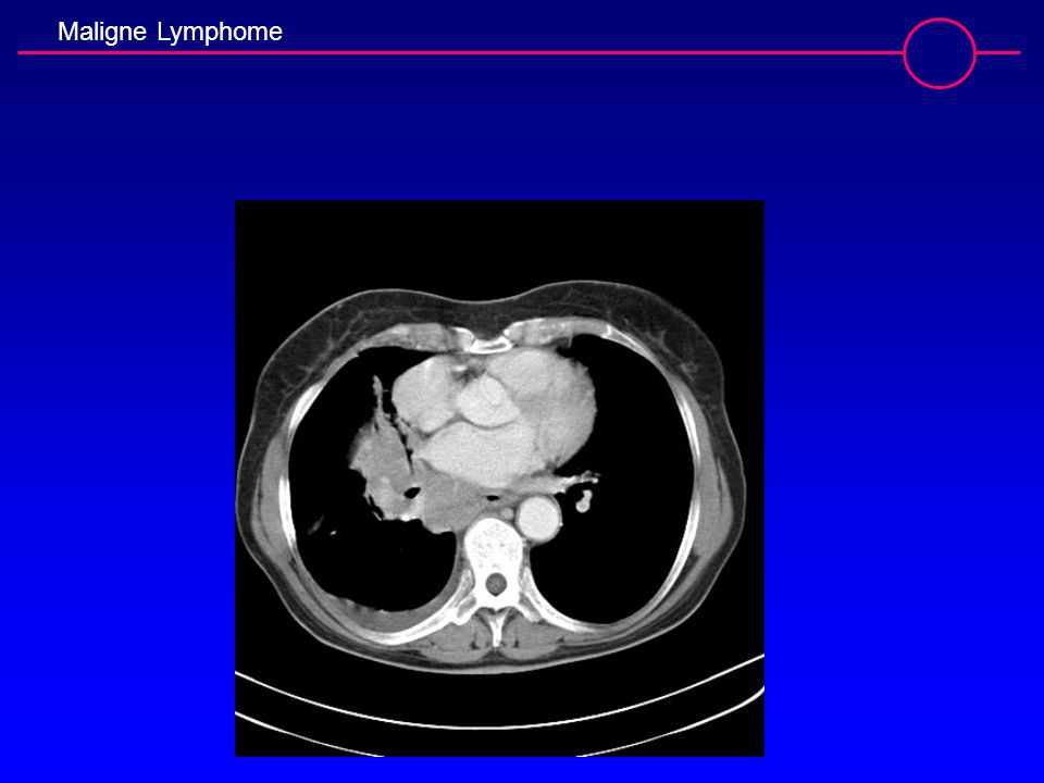
Kasuistiken 1. Patientin mit malignem Lymphom, Alter bei ED: 63 Jahre
Malignes Lymphom, ED 03/2005 Lokalisation: Ausgedehnte mediastinale Lymphome mit Ausmauerung des gesamten Mediastinums (größter ø 14 x 10 cm), poststenotische pulmonale Infiltrate re. Sowie klein nodaler pulmonaler Befall bds., Pleuraerguss re., supraclaviculäre Lymphome bds. Z.n. CT-gesteuerter Biopsie Histologie: Diffus großzelliges Non-Hodgkin-Lymphom, Stadium IVB
, poststenotische pulmonale Infiltrate re. Sowie klein nodaler pulmonaler Befall bds., Pleuraerguss re., supraclaviculäre Lymphome bds. Z.n. CT-gesteuerter Biopsie. Histologie: Diffus großzelliges Non-Hodgkin-Lymphom, Stadium IVB ")
25
Kasuistiken 1. Patientin mit malignem Lymphom ( Fortsetzung)
Z.n. Vorphase-Chemotherapie mit Vincristin und Steroiden 03/2005 Z.n. 8 Zyklen R-CHOP-Chemoimmunotherapie 03-08/2005 Stand 09/2005: Komplette Remission Stand 09/2006: Anhaltende komplette Remission

26
Wichtigste Grundlage für die Behandlung ist die feingewebliche Diagnosesicherung:
Bei Diagnose CT-gesteuerte Biopsie Nach 4 x R-CHOP-Therapie

28
Kasuistiken 2. Patient mit Mantelzell-Lymphom, Alter bei ED: 56 Jahre
ED 01/95, Stadium: II (fraglich IV) Z.n. 8 x COP 01/95 – 07/95 07/96: Histologisch gesicherte Manifestation Dickdarm- und Rektumschleimhaut sowie Sinus piriformis Z.n. 1 x CHEP 08/96 Z.n. 2 x CHEP 09/96 : Komplette Remission Z.n. Hochdosis-Chemotherapie (BEAM) mit autologer peripherer Stammzelltransplantation
 Z.n. 8 x COP 01/95 – 07/95. 07/96: Histologisch gesicherte Manifestation Dickdarm- und Rektumschleimhaut sowie Sinus piriformis. Z.n. 1 x CHEP 08/96. Z.n. 2 x CHEP 09/96 : Komplette Remission. Z.n. Hochdosis-Chemotherapie (BEAM) mit autologer peripherer Stammzelltransplantation ")
29
Kasuistiken 2. Patient: Mantelzell-Lymphom (Fortsetzung)
04/2002: Anhaltende komplette Remission 10/2002: Hochgradiger V.a. Rezidiv im Bereich des Waldeyer´schen Rachenrings Z.n. Biopsie und Laserresektion 12/2002 Histologieergebnis : Malignes B-Zell-Lymphom entsprechend einem Mantelzell-Lymphom Z.n. 6. R-CHP 01-05/2003: Komplette Remission 09/2006: Anhaltende komplette Remission

30
Niedrigmalignes (indolentes) Lymphom
 Lymphom")
31
Maligne Lymphome - Heilungschancen
KEINE HEILUNG (wenige können geheilt werden) CLL
 CLL.")
32
B-CLL: Stadieneinteilung und Prognose
Rai Charakteristika Medianes Risiko Überleben 0 Lymphozytose in PB und KM > 120 Monate Niedrig I Lymphozytose u. vergrößerte LK Monate Mittel II Lymphozytose u. Splenomegalie Monate Mittel oder Hepatomegalie III Lymphozytose u. Hb < 11g/dl Monate Hoch IV Lymphozytose u. Plt < /µl Monate Hoch

33
B-CLL: Stadieneinteilung und Prognose
Binet Charakteristika Medianes Risiko Überleben A` Lymphozyten < / µl > 120 Monate Niedrig + Hb > 12 g/dl A`` Lymphozyten < / µl Monate Mittel + Hb < 12 g/dl B Hb < 10 g/dl; Plt > /µl Monate Mittel > 3 vergrößerte Lymphknotenstationen C Hb < 10 g/dl; Plt < /µl Monate Hoch (Bei Stadium A dürfen nicht mehr als 2 Lymphknotenstationen befallen sein, bei Stadium C spielt die Anzahl der befallenen Lymphknotenstationen keine Rolle.)
")
34
B-CLL - Behandlungsprinzipien
Binet A + Binet B ohne Symptome Keine Behandlung Binet B mit Symptomen + Binet C Behandlung

35
Maligne Lymphome - Heilungschancen
Niedrigmaligne (indolent) Stadium III + IV Stadium I + II HEILUNG KEINE HEILUNG (wenige können geheilt werden)
 Stadium III + IV. Stadium I + II. HEILUNG. KEINE HEILUNG. (wenige können geheilt werden)")
36
Niedrigmaligne (indolente) Lymphome - Behandlungsprinzipien -
Stadium I + Stadium II Strahlentherapie Stadium III + Stadium IV mit Symptomen ohne Symptome Behandlung Beobachtung Strahlen- therapie Chemoimmuno- therapie Radioimmuno- therapie Hochdosis- therapie

37
Niedrigmaligne (indolente) Lymphome Follicular lymphoma international prognostic index F L I P I
Alter > 60 Jahre Stadium III oder IV Hb < 120 g/L Mehr als 4 befallene Lymphknotenareale Serum LDH erhöht Für jedes Charakteristikum wird 1 Punkt vergeben: Punkte Risikogruppe Jahresüberleben Niedrig % Intermediär % Hoch %

38
Kasuistiken 3. Patient mit CBCC, Alter bei ED: 59 Jahre
CBCC, ED 1990, Stadium IV A Vortherapie mit PROMACE, CHOP, Ifosphamid/Steroide seit 1995 massiver Hautbefall seit 05/98 Therapie refraktär 06/98: Z.n. 1x Fludarabintherapie (kein Ansprechen) 07/98: 4x Rituximab (PR) 01/99: Progress 02/99: 4x Rituximab (kein Ansprechen) 03-04/99: 1x Bendamustin/Mitoxantron/Rituximab (2x) 1x Bendamustin/Mitoxantron seither CR, 120+ Monate
 07/98: 4x Rituximab (PR) 01/99: Progress. 02/99: 4x Rituximab (kein Ansprechen) 03-04/99: 1x Bendamustin/Mitoxantron/Rituximab (2x) 1x Bendamustin/Mitoxantron. seither CR, 120+ Monate.")
39
Exulzeriertes Hautlymphom vor Therapie

40
Exulzeriertes Hautlymphom 4 Wochen nach BMR

41
Exulzeriertes Hautlymphom 8 Wochen nach BMR

42
Kasuistiken 4. Patientin mit Non-Hodgkin-Lymphom, Alter bei ED: 60 Jahre ED 12/2001, initiales Stadium IV B Histologie: Follikuläres Lymphom Grad I Initialbefall: Lymphome submandibulär/cervical/claviculär li., axillär bds., paraaortal und parailiacal re., inguinal bds., Knochenmarkbefall, Nachtschweiß als B-Symptomatik Z.n. Polychemotherapie (6 x CHOP) 01-05/2002 06/2002: Komplette Remission
 01-05/ /2002: Komplette Remission ")
43
Kasuistiken 4. Patientin mit Non-Hodgkin-Lymphom (Fortsetzung)
Progress 11/2002: Multiple subcutane Lymphombildungen Histologie:Follikuläres Non-Hodgkin-Lymphom, Grad IIIA Z.n. 1x Bendamustin/Mitoxantrone/ Rituximab, 3 x Bendamustin/Mitoxantrone, 12/ /2003 Staging in 05/2003: Komplette Remission des Nodalbefalls, neu aufgetretene Raumforderungen pulmonal

44
Kasuistiken 4. Patientin mit Non-Hodgkin-Lymphom (Fortsetzung)
Z.n. Fluconazol-Therapie bei V.a. oligosymptomatische Pilzpneumonie 08-09/2003 Staging in 12/2003: Status idem Progress 06/2004: Lokalrezidiv Unterschenkel li. Z.n. Strahlentherapie GHD 30 Gy, ED 3 Gy 07-08/2004 Progress 10/2004: Lymphombildung inguinal re. Symptomatischer Progress 03/2005: Weiter progrediente Lymphknotenbildung inguinal re.

45
Kasuistiken 4. Patientin mit Non-Hodgkin-Lymphom (Fortsetzung)
Z.n. Chemoimmunotherapie (3 Zyklen Bendamustin/ Mitoxantrone/Rituximab) 04-06/2005 Staging 07/2005: Komplette Remission Z.n. weiterer Radioimmuntherapie (2 Einzelgaben Rituximab gefolgt von Zevalin) 08/2005 Staging 10/2006: Anhaltende komplette Remission
 04-06/2005. Staging 07/2005: Komplette Remission. Z.n. weiterer Radioimmuntherapie (2 Einzelgaben Rituximab gefolgt von Zevalin) 08/2005. Staging 10/2006: Anhaltende komplette Remission.")
47
Kasuistiken 5. Patientin mit kleinzelligem B-Zell-Lymphom,
ED 06/2000, Stadium IV B (Initialbefall bds. cervical, bds. axillär, Knochenmark) Zustand nach 6 Zyklen R-CHOP/Chemoimmunotherapie 07-10/00: Komplette Remission Stand 10/06: Anhaltende komplette Remission 5. Patientin mit kleinzelligem B-Zell-Lymphom, Alter bei ED: 47 Jahre
 Zustand nach 6 Zyklen R-CHOP/Chemoimmunotherapie 07-10/00: Komplette Remission. Stand 10/06: Anhaltende komplette Remission. 5. Patientin mit kleinzelligem B-Zell-Lymphom, Alter bei ED: 47 Jahre.")
Ähnliche Präsentationen


>")


>")


