Präsentation herunterladen
Die Präsentation wird geladen. Bitte warten

1
6.5 Eiweiße = Proteine

2
Wo finden wir Proteine?

3
Haare Figure :3-10 part a Title: Structural proteins part a Hair
Caption: Common structural proteins include the keratin of (a) hair and (b) horn, and (c) the silk protein in a spider web. Haare
 hair and (b) horn, and (c) the silk protein in a spider web. Haare.")
4
Horn Figure :3-10 part b Title: Structural proteins part b Horn
Caption: Common structural proteins include the keratin of (a) hair and (b) horn, and (c) the silk protein in a spider web. Horn
 hair and (b) horn, and (c) the silk protein in a spider web. Horn.")
5
Seide Figure :3-10 part c Title: Structural proteins part c Silk
Caption: Common structural proteins include the keratin of (a) hair and (b) horn, and (c) the silk protein in a spider web. Seide
 hair and (b) horn, and (c) the silk protein in a spider web. Seide.")
6
Wie sind Proteine strukturiert?

7
Aminosäure Dipeptid Peptid bindung Wasser Figure :3-12 Title:
Protein synthesis Caption: In protein synthesis, a dehydration synthesis joins the carbon of the carboxyl acid group of one amino acid to the nitrogen of the amino group of a second amino acid. The resulting covalent bond is called a peptide bond.

8
Sekundärstruktur Tertiärstruktur Quartärstruktur Primärstruktur
gly leu leu val val Tertiärstruktur lys lys lys gly his lys ala gly lys val his lys pro ala lys val lys Figure :3-13 Title: The four levels of protein structure Caption: Levels of protein structure are represented here by hemoglobin, the oxygen-carrying protein in red blood cells (the red discs represent the iron-containing heme group that binds oxygen). All levels of protein structure are determined by the amino acid sequence of the protein, interactions among the R groups of the amino acids, and interactions between the R groups and their surroundings. Question Why do most proteins, when heated, lose their ability to function? pro Quartärstruktur Primärstruktur
. All levels of protein structure are determined by the amino acid sequence of the protein, interactions among the R groups of the amino acids, and interactions between the R groups and their surroundings. Question Why do most proteins, when heated, lose their ability to function pro. Quartärstruktur. Primärstruktur.")
9
Reihenfolge der Aminosäuren, mit Peptidbindungen verbunden
leu val lys Reihenfolge der Aminosäuren, mit Peptidbindungen verbunden lys gly his pro ala lys val lys Figure :3-13 Title: The four levels of protein structure Caption: Levels of protein structure are represented here by hemoglobin, the oxygen-carrying protein in red blood cells (the red discs represent the iron-containing heme group that binds oxygen). All levels of protein structure are determined by the amino acid sequence of the protein, interactions among the R groups of the amino acids, and interactions between the R groups and their surroundings. Question Why do most proteins, when heated, lose their ability to function? pro Primärstruktur
. All levels of protein structure are determined by the amino acid sequence of the protein, interactions among the R groups of the amino acids, and interactions between the R groups and their surroundings. Question Why do most proteins, when heated, lose their ability to function pro. Primärstruktur.")
10
Helix oder β-Faltblatt durch H-Brücken zwischen Aminosöuren
Sekundärstruktur Helix oder β-Faltblatt durch H-Brücken zwischen Aminosöuren

11
Viele Helices und Faltblatt-Strukturen dreidimensional zusammen,
Tertiärstruktur Viele Helices und Faltblatt-Strukturen dreidimensional zusammen, durch Ionenbindungen und Disulfidbrücken Figure :3-13 Title: The four levels of protein structure Caption: Levels of protein structure are represented here by hemoglobin, the oxygen-carrying protein in red blood cells (the red discs represent the iron-containing heme group that binds oxygen). All levels of protein structure are determined by the amino acid sequence of the protein, interactions among the R groups of the amino acids, and interactions between the R groups and their surroundings. Question Why do most proteins, when heated, lose their ability to function? pro
. All levels of protein structure are determined by the amino acid sequence of the protein, interactions among the R groups of the amino acids, and interactions between the R groups and their surroundings. Question Why do most proteins, when heated, lose their ability to function pro.")
12
Einige Polypeptide zusammen
Prosthetische Gruppe (ein nicht-Protein-Teil) Konjugiertes Protein Quartärstruktur
 Konjugiertes Protein. Quartärstruktur.")
13
Polare und nichtpolare Aminosäuren
hydrophile Restgruppen nichtpolare Aminosäuren hydrophobe Restgruppen

14
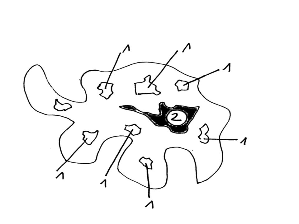
Damit hydrophile Substanzen durch eine Membran gehen können nutzen sie Protein-Kanäle (1). Diese besitzen polare Aminosäuren, die hydrophil sind. Wenn die Kanäle durch negativ geladene Restgruppen gebildet werden, können positiv geladene Ionen diffundieren und umgekehrt.

15
Polare (hydrophile) Aminosäuren
Wichtig für den Transport hydrophiler Stoffe

16
Nicht-Polare (hydrophobe) Aminosäuren
Wichtig für Verankerung in der Membran

18
Unterschied Faser- und Kugelprotein

19
Kollagen, Aktin und Myosin sind Faserproteine

20
Faserproteine sind lang gestreckt und schmal!

21
Faserproteine sind meist wasserunlöslich!

22
Faserproteine sind in Haaren, Horn, Muskeln und Haut
Ihre Funktion ist meist, dass sie etwas sind.

23
Kugelprotein z.B. Insulin
Sind rund und wasserlöslich! Ihre Funktion ist meist, dass sie etwas tun: ENZYME

24
Faserprotein Kugelprotein Löslichkeit Form Funktion Beispiel

25
Faserprotein Kugelprotein Wasserunlöslich Wasserlöslich Form Funktion
Löslichkeit Wasserunlöslich Wasserlöslich Form Funktion Beispiel

26
Faserprotein Kugelprotein Löslichkeit Wasserunlöslich Wasserlöslich Form Funktion Beispiel

27
Faserprotein Kugelprotein Löslichkeit Wasserunlöslich Wasserlöslich Form Lang gestreckt und schmal Eng gefaltet dadurch rund Funktion Beispiel

28
Faserprotein Kugelprotein Löslichkeit Wasserunlöslich Wasserlöslich Form Lang gestreckt und schmal Eng gefaltet dadurch rund Funktion Beispiel

29
Faserprotein Kugelprotein Löslichkeit Wasserunlöslich Wasserlöslich Form Lang gestreckt und schmal Eng gefaltet dadurch rund Funktion Bilden meist Strukturen Sie tun meist etwas Beispiel

30
Faserprotein Kugelprotein Löslichkeit Wasserunlöslich Wasserlöslich Form Lang gestreckt und schmal Eng gefaltet dadurch rund Funktion Bilden meist Strukturen Sie tun meist etwas Beispiel

31
Faserprotein Kugelprotein Löslichkeit Wasserunlöslich Wasserlöslich Form Lang gestreckt und schmal Eng gefaltet dadurch rund Funktion Bilden meist Strukturen Sie tun meist etwas Beispiel Kollagen, Myosin, Keratin Hämoglobin, Hormone

32
Funktionen von Proteinen und ein Beispiel
Funktion: Enzym Beispiel: Katalase Form: Kugelprotein

33
Funktionen von Proteinen und ein Beispiel
Funktion: Struktur Beispiel: Kollagen Form: Faserprotein

34
Funktionen von Proteinen und ein Beispiel
Funktion: Transport Beispiel: Hämoglobin Form: Kugelprotein

35
Funktionen von Proteinen und ein Beispiel
Funktion: Bewegung Beispiel: Myosin Form: Faserprotein

36
Funktionen von Proteinen und ein Beispiel
Funktion: Hormon Beispiel: Insulin Form: Kugelprotein

37
Funktionen von Proteinen und ein Beispiel
Funktion: Abwehr Beispiel: Immunoglobulin Form: Kugelprotein

Ähnliche Präsentationen


>")


