Präsentation herunterladen
Die Präsentation wird geladen. Bitte warten

1
Glutamate and GABA levels in the frontal cortex of rats with chronic epilepsy. H. Baran, P. Kalina, J. Wallner, H. Papst and B. Kepplinger Rev. Bras. Neurol. (2006) Karl Landsteiner Institut LKM Amstetten-Mauer Biochemische Grundlagen der Neurophysiologie, Department der Naturwissenschaften, VM Universität Wien Neurologische Abteilung LKM Amstetten-Mauer
 Karl Landsteiner Institut LKM Amstetten-Mauer Biochemische Grundlagen der Neurophysiologie, Department der Naturwissenschaften, VM Universität Wien Neurologische Abteilung LKM Amstetten-Mauer.")
2
Chronische Epilepsie ca. 70 % der Patienten sind gut einstellbar… ca 20 % weisen unter einer antikonvulsiven Therapie weiterhin gehäufte Anfälle auf…. und ca 10 % sind weitestgehend therapieresistent. Gibt es ein Tiermodell, an dem man das Phänomen der chronischen Epilepsie mit unterschiedlicher klinischer Ausprägung studieren kann - um die molekularbiologisch Vorgänge im Gehirn besser verstehen zu können und damit neue pharmakologische Strategien entwickeln zu können?

3
Endogene und exogene exzitatorische Aminosäuren spielen eine wesentliche Rolle bei der Epilepsie

4
Kainsäure ist ein Agonist eines glutamatergen exzitatorischen Aminosäure-Rezeptor- Subtyps
GLUTAMATE RECEPTORS NMDA AMPA KAINATE METABOTROPIC Glutamate Site Glycine Site Agonists: NMDA Glycine Quisqualic acid Kainic acid L-AP4 D-Serin AMPA Domoic acid ACPD R-HA966 Selective Antagonists: KYNA KYNA KYNA KYNA MCPG AP Cl-KYNA NBQX CNQX AP ,7-Cl-KYNA GYKI 52466 CGS MNQX CGP L-689,560 CPP Effector pathways: NA+/K+/Ca NA+/K+/Ca NA+/K+/Ca IP3DAG J. R. Cooper et al., 1996, Biochemical Basis of Neuropharmacology, Oxford University Press (simplified).
.")
5
Kainsäure Epilepsie Modell
The behavioural alterations in rats and the neurochemical and histopathological changes in rat brains after systemic kainic acid (KA) application show similarities to the behavioural, neurochemical and histopathological alterations observed in human temporal lobe epilepsy. (Olney et al., 1974; Nadler, 1981; Sperk 1983; Baran, 1985; Ben-Ari, 1985; Meldrum 1991; Löscher 1993; Baran, 2004) .
 application show similarities to the behavioural, neurochemical and histopathological alterations observed in human temporal lobe epilepsy. (Olney et al., 1974; Nadler, 1981; Sperk 1983; Baran, 1985; Ben-Ari, 1985; Meldrum 1991; Löscher 1993; Baran, 2004) .")
6
KA-Modell für die chronische Epilepsie
KA-treated rats during 6 months after KA administration (10 mg/kg, s.c.).
.")
7
Glutamat und GABA

8
Glutamate decarboxylase (GAD) activity in KA-rats with spontaneous seizures (rating = 3 ÷ 4)
Baran et al., 2004, Neurosignals

9
Glutamate and GABA in brain regions of KA-rats with spontaneous seizures (rating = 3 ÷ 4)
")
10
GABA/Glutamate ratio

11
Die Ergebnisse zeigen beim chronischen KA-Epilepsiemodell
Befunde Die Ergebnisse zeigen beim chronischen KA-Epilepsiemodell ein charakteristisches Muster der erhöhten GABA-ergen Aktivität in verschiedenen Hirnregionen. Dies läßt vermuten, daß die veränderte GABA-erge Aktivität eine funktionale Rolle in einem “neu ausgebildeten neuronalen Netzwerk” aufweist und die spontanen repetitiven Anfälle verursacht.

12
Resumè An diesem Modell soll künftig geprüft werden, ob
GABA-mimetische Medikamente oder Substanzen, die eine NMDA Rezeptor-mediierte Freisetzung von GABA bewirken eine antiepileptische und neuroprotektive Wirkung zeigen.

13
Vielen Dank für Ihre Aufmerksamkeit !

14
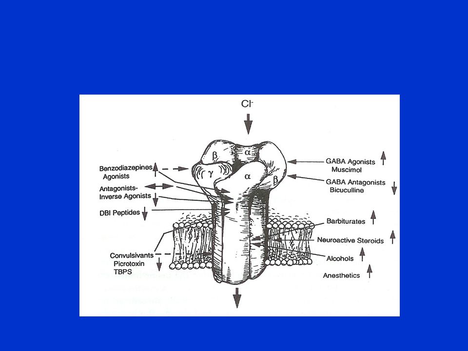
Modulation of GABAergic activity

16
6 months after KA injection we analyzed
The levels of glutamate and GABA in different brain regions and The activity of glutamic acid decarboxylase (GAD) in different brain regions.
 in different brain regions.")
Ähnliche Präsentationen


>")


![Nernst Gleichung: Wird das Potential negativer oder positiver wenn die Konzentration von [X]i zunimmt? Welches Potential hat man wenn [X]i = [X]o ist?](/1/650474/big_thumb.jpg "Nernst Gleichung: Wird das Potential negativer oder positiver wenn die Konzentration von [X]i zunimmt? Welches Potential hat man wenn [X]i = [X]o ist?>")
 61 Fe reaction at the TRIGA reactor in Mainz and future possibilities at FRANZ T. Heftrich 1, M. Mikorski 1, C. Beinrucker.>")
 4th Planet Formation Workshop MPIA, 1 st March 2006 Discovery of a cool 5.5 Earth-mass planet through gravitational.>")

