Präsentation herunterladen
Die Präsentation wird geladen. Bitte warten

1
Moderne nicht-invasive Methoden zur Erforschung des menschlichen Gehirns
Priv.-Doz. Dr. Carsten Wolters Dr.rer.nat. Harald Kugel Dr.med. Gabriel Möddel Priv.Doz. Dr. med. Christoph Kellinghaus Tensor weiter erlaeutern Zeitableitung Programmlaenge, 1Mill. WM-Rendering->5000 nodes 1:10 raus und anfangs erwaehnen Vorlesung, 15.Oktober 2013

2
Outline General planning for this lecture (language? date/time? required knowledge? Participants- -List!) Literature for this lecture Introduction to the lecture (Part 1)
")
3
Aktuelle Vorlesungsplanung
15.Oktober: Vorbesprechung und Motivation (Wolters) 22.Oktober: Einführung Magnetresonanztomographie (MRT) (Kugel) 29.Oktober: Medizinische Grundlagen zur Elektro- (EEG) und Magnetoencephalography (MEG) (Wolters) 5.Nov.: Mathematisch-physikalische Modellierungsgrundlagen zu EEG und MEG, Teil 1 (Wolters) 12.Nov.: Mathematisch-physikalische Modellierungsgrundlagen zu EEG und MEG, Teil 2 (Wolters) 19.Nov.: Grundlagen von Epilepsie und EEG (Kellinghaus) 26.Nov.: Epileptische Anfälle und ihre Behandlung (Kellinghaus) 3.Dez.: Registrierung von MRT: Teil 1 (Wolters) 10.Dez3.: Registrierung von MRT: Teil 2 (Wolters) 17.Dez.: Segmentierung von MRT (Wolters) 7.Jan.: Mathematik des EEG/MEG Vorwärtsproblems, Teil 1 (Wolters) 14.Jan.: Mathematik des EEG/MEG Vorwärtsproblems, Teil 2 (Wolters) 21.Jan.: Mathematik des EEG/MEG inversen Problems, Teil 1 (Wolters) 28.Jan.: Mathematik des EEG/MEG inversen Problems, Teil 2 (Wolters) 4.Feb.: Epilepsiechirurgie, Teil 3 (Möddel)
 22.Oktober: Einführung Magnetresonanztomographie (MRT) (Kugel) 29.Oktober: Medizinische Grundlagen zur Elektro- (EEG) und Magnetoencephalography (MEG) (Wolters) 5.Nov.: Mathematisch-physikalische Modellierungsgrundlagen zu EEG und MEG, Teil 1 (Wolters) 12.Nov.: Mathematisch-physikalische Modellierungsgrundlagen zu EEG und MEG, Teil 2 (Wolters) 19.Nov.: Grundlagen von Epilepsie und EEG (Kellinghaus) 26.Nov.: Epileptische Anfälle und ihre Behandlung (Kellinghaus) 3.Dez.: Registrierung von MRT: Teil 1 (Wolters) 10.Dez3.: Registrierung von MRT: Teil 2 (Wolters) 17.Dez.: Segmentierung von MRT (Wolters) 7.Jan.: Mathematik des EEG/MEG Vorwärtsproblems, Teil 1 (Wolters) 14.Jan.: Mathematik des EEG/MEG Vorwärtsproblems, Teil 2 (Wolters) 21.Jan.: Mathematik des EEG/MEG inversen Problems, Teil 1 (Wolters) 28.Jan.: Mathematik des EEG/MEG inversen Problems, Teil 2 (Wolters) 4.Feb.: Epilepsiechirurgie, Teil 3 (Möddel)")
4
Outline Literature for this lecture
Introduction to the lecture (Part 1)
")
5
Literature for this lecture
Lecture webside: PEBBLES: Parallel and Element Based grey Box Linear Equation Solver

6
Outline Literature for this lecture
Introduction to the lecture (Part 1)
")
7
Basics of clinical EEG and MEG

8
Electro- (EEG) and Magneto-encephalography (MEG)
275 channel axial gradiometer whole-cortex MEG 128 channel EEG Cohen et al. measured first MEG in 1968: alpha-activity is about 10^7 times weaker than the earth magnetic field

9
Spatial and temporal resolution of brain imaging methods
[Gazzaniga, Ivry & Mangun, Cognitive Neuroscience, 2nd ed., W.W.Norton & Company, 2002] Spatial and temporal resolution of brain imaging methods

10
Grundlagen klinischer EEG und MEG Anwendungen
EEG ist Standard in der klinischen Praxis MEG ist kostenintensiv (Gerätekosten, Wartung, Heliumkühlung…) Datenauswertung ist komplex (wie auch für EEG, fMRT, …) In Deutschland bisher keine Vergütung durch die Krankenkassen => Warum also MEG?
 Datenauswertung ist komplex (wie auch für EEG, fMRT, …) In Deutschland bisher keine Vergütung durch die Krankenkassen. => Warum also MEG")
11
Grundlagen - MEG MEG registriert nicht-invasiv magnetische Felder neuronaler Aktivität Ähnlich dem EEG: Ableitung neuronaler Aktivität MEG und EEG messen Aktivität derselben Generatoren PET oder fMRT: Indirekte Erfassung neuronaler Aktivität 4D Neuroimaging, San Diego, CA, USA

12
Magnetische Abschirmkammer

13
MEG Interna

14
Erfassung des magnetischen Flusses
Magnetometer Superconducting quantum interference device (SQUID) Axiales Gradiometer Planares Gradiometer Papanicolaou (Ed.): Clinical Magnetoencephalography and Magnetic Source Imaging
 Axiales Gradiometer. Planares Gradiometer. Papanicolaou (Ed.): Clinical Magnetoencephalography and Magnetic Source Imaging.")
15
MEG-System am IBB, Uni Münster
[Lanfer, diploma thesis, 2007] MEG-System am IBB, Uni Münster Finite Elemente Knoten für die MEG Sensor-Beschreibung 15

16
Epileptic activity as measured with EEG and MEG

17
Source analysis in presurgical epilepsy diagnosis
0.5%-1% of world population suffers from epilepsy 70-80% of patients successfully treated with drugs For those who are still pharma-resistent after 2-3 drugs Probability of success of a further different drug: 6% (Wiebe et al 2001) Probability of success of a surgical treatment: 50% (Wiebe et al 2001) Indispensable prerequisite for surgery: Focal epilepsy->Localization Gold standard: Video-monitoring and visual inspection of the EEG (Wilson 1996) MRI: Identification of an underlying lesion PET and Neuropsychology: Localization of a functional deficit Source analysis of EEG seizure (ictal) activity (Plummer et al., 2008) EEG/MEG interictal activity: “irritative zone” (Stefan et. al., 2003) PEBBLES: Parallel and Element Based grey Box Linear Equation Solver
 Probability of success of a surgical treatment: 50% (Wiebe et al 2001) Indispensable prerequisite for surgery: Focal epilepsy->Localization. Gold standard: Video-monitoring and visual inspection of the EEG (Wilson 1996) MRI: Identification of an underlying lesion. PET and Neuropsychology: Localization of a functional deficit. Source analysis of. EEG seizure (ictal) activity (Plummer et al., 2008) EEG/MEG interictal activity: irritative zone (Stefan et. al., 2003) PEBBLES: Parallel and Element Based grey Box Linear Equation Solver.")
18
Epileptic spikes in EEG and MEG
Clear spike in EEG Nearly no/no signal in MEG

19
Epileptic spikes in EEG and MEG
Clear spike in EEG Nearly no/no signal in MEG Deep source Strongly radially oriented source

20
Sensitivity for radial and tangential sources
MEG registers mainly tangential source components: Sulci-walls: tangential pyramidal cells -> High amplitudes „Diagonal“ orientation -> Medium amplitude Radial sources hardly produce an MEG: Depth and crown of sulci: radial pyramidal cells -> Low contribution

21
Epileptic spikes in EEG and MEG
Clear signal in MEG, poor signal in EEG Explanation?

22
Sensitivity Sensitivity EEG > MEG in deep areas
But: Sensitivity MEG > EEG in superficial areas Goldenholz et al., 2009

23
Spikes in EEG and MEG What should we use?
MEG instead of EEG? Only EEG? Seite 23 Iwasaki et al., 2005

24
Combined EEG and MEG 275 channel axial gradiometer whole-cortex MEG
128 channel EEG Cohen et al. measured first MEG in 1968: alpha-activity is about 10^7 times weaker than the earth magnetic field

25
Source analysis of interictal spikes in presurgical epilepsy diagnosis

26
Averaged interictal EEG spikes
[Wolters & Kellinghaus, 2006] Averaged interictal EEG spikes About 0.25% of the world population suffers from drug-resistnt epilepsy and bout 10-15% would profit from a surgical removl of the epileptogenic tissue. 42 year old female suffering from medically intractable complex partial seizures of mesial-temporal origin Seizures had duration of up to 10 minutes and a frequency of twice to 5 times a month Measurements: ========= A short-term barbiturate (Schlafmittel), known to provoke epileptiform activity -> 60-channel EEG montage with reference at the ear contralateral to the epileptic focus Source reconstruction: ============== Single dipole fit at signal peak ->Localization of the secondary activations, propagating along the temporal cortical surface ->Primary activity in the hippocampus and the amygdala (cannot be measured outside) Therapy: ====== Selective amygdalohippocampectomy -> Patient free of seizures Measure EEG and/or MEG
, known to provoke epileptiform activity. -> 60-channel EEG montage with reference at the ear contralateral to the epileptic focus. Source reconstruction: ============== Single dipole fit at signal peak. ->Localization of the secondary activations, propagating along the temporal cortical surface. ->Primary activity in the hippocampus and the amygdala (cannot be measured outside) Therapy: ====== Selective amygdalohippocampectomy. -> Patient free of seizures. Measure EEG and/or MEG.")
27
Results of combined EEG/MEG dipole fit
[Wolters & Kellinghaus, 2006] Results of combined EEG/MEG dipole fit EEG data and (transparent) cortex MEG data and (transparent) cortex 42 year old female suffering from medically intractable complex partial seizures of mesial-temporal origin Seizures had duration of up to 10 minutes and a frequency of twice to 5 times a month Measurements: ========= A short-term barbiturate (Schlafmittel), known to provoke epileptiform activity -> 60-channel EEG montage with reference at the ear contralateral to the epileptic focus Source reconstruction: ============== Single dipole fit at signal peak ->Localization of the secondary activations, propagating along the temporal cortical surface ->Primary activity in the hippocampus and the amygdala (cannot be measured outside) Therapy: ====== Selective amygdalohippocampectomy -> Patient free of seizures Inverse method: Single current dipole
 cortex. MEG data and (transparent) cortex. 42 year old female suffering from medically intractable complex partial seizures of mesial-temporal origin. Seizures had duration of up to 10 minutes and a frequency of twice to 5 times a month. Measurements: ========= A short-term barbiturate (Schlafmittel), known to provoke epileptiform activity. -> 60-channel EEG montage with reference at the ear contralateral to the epileptic focus. Source reconstruction: ============== Single dipole fit at signal peak. ->Localization of the secondary activations, propagating along the temporal cortical surface. ->Primary activity in the hippocampus and the amygdala (cannot be measured outside) Therapy: ====== Selective amygdalohippocampectomy. -> Patient free of seizures. Inverse method: Single current dipole.")
28
Results of combined EEG/MEG L1 norm current density reconstruction
[Wolters & Kellinghaus, 2006] Results of combined EEG/MEG L1 norm current density reconstruction EEG data and (nontransparent) cortex MEG data and (nontransparent) cortex 42 year old female suffering from medically intractable complex partial seizures of mesial-temporal origin Seizures had duration of up to 10 minutes and a frequency of twice to 5 times a month Measurements: ========= A short-term barbiturate (Schlafmittel), known to provoke epileptiform activity -> 60-channel EEG montage with reference at the ear contralateral to the epileptic focus Source reconstruction: ============== Single dipole fit at signal peak ->Localization of the secondary activations, propagating along the temporal cortical surface ->Primary activity in the hippocampus and the amygdala (cannot be measured outside) Therapy: ====== Selective amygdalohippocampectomy -> Patient free of seizures Inverse method: L1 norm current density
 cortex. MEG data and (nontransparent) cortex. 42 year old female suffering from medically intractable complex partial seizures of mesial-temporal origin. Seizures had duration of up to 10 minutes and a frequency of twice to 5 times a month. Measurements: ========= A short-term barbiturate (Schlafmittel), known to provoke epileptiform activity. -> 60-channel EEG montage with reference at the ear contralateral to the epileptic focus. Source reconstruction: ============== Single dipole fit at signal peak. ->Localization of the secondary activations, propagating along the temporal cortical surface. ->Primary activity in the hippocampus and the amygdala (cannot be measured outside) Therapy: ====== Selective amygdalohippocampectomy. -> Patient free of seizures. Inverse method: L1 norm current density.")
29
Source analysis of seizure (ictal) spikes in presurgical epilepsy diagnosis
 spikes in presurgical epilepsy diagnosis")
30
Typical EEG signals Gamma(30-70Hz): Starke Konzentr., Lernphase
[Gazzaniga, Ivry & Mangun, Cognitive Neuroscience, 2nd ed., W.W.Norton & Company, 2002] Typical EEG signals Gamma(30-70Hz): Starke Konzentr., Lernphase Beta (14-30Hz): Hellwach, gute Intelligenzleistung Alpha (8-13Hz): Entspannte Wachheit Theta (4-7Hz): Leichte Schlafphasen Delta ( Hz): Traumlose Tiefschlafphase drowsy=schlaefrig
: Starke Konzentr., Lernphase. Beta (14-30Hz): Hellwach, gute Intelligenzleistung. Alpha (8-13Hz): Entspannte Wachheit. Theta (4-7Hz): Leichte Schlafphasen. Delta ( Hz): Traumlose Tiefschlafphase. drowsy=schlaefrig.")
31
EEG Preprocessing Nächste Folie kommen Ergebnisse!
[Rullmann, Anwander, Dannhauer, Warfield, Duffy & Wolters, NeuroImage, 44(2), 2009] Nächste Folie kommen Ergebnisse! 31
, 2009] Nächste Folie kommen Ergebnisse! 31.")
32
T1 MRI segmentation Segmentierung per Hand angepasst
[Rullmann, Anwander, Dannhauer, Warfield, Duffy & Wolters, NeuroImage, 44(2), 2009] T1 MRI segmentation Segmentierung per Hand angepasst Vergitterung: jedes gewebe eigenes Label Eogentliche gitter ist feiner (1mm) hier nur 3mm dargestellt
, 2009] T1 MRI segmentation. Segmentierung per Hand angepasst. Vergitterung: jedes gewebe eigenes Label. Eogentliche gitter ist feiner (1mm) hier nur 3mm dargestellt.")
33
FE mesh generation Segmentierung per Hand angepasst
[Rullmann, Anwander, Dannhauer, Warfield, Duffy & Wolters, NeuroImage, 44(2), 2009] Segmentierung per Hand angepasst Vergitterung: jedes gewebe eigenes Label Eogentliche gitter ist feiner (1mm) hier nur 3mm dargestellt
, 2009] Segmentierung per Hand angepasst. Vergitterung: jedes gewebe eigenes Label. Eogentliche gitter ist feiner (1mm) hier nur 3mm dargestellt.")
34
Brain conductivity anisotropy modeling
[Rullmann, Anwander, Dannhauer, Warfield, Duffy & Wolters, NeuroImage, 44(2), 2009] Brain conductivity anisotropy modeling Original DTI data FA map after registration FA map on T1-MRI Effective medium approach model (DTI <-> CTI): Model DTI<->Conductivity Tensor Image (CTI) [Tuch et al., Ann. NYAS, 1999] Linear model DTI<->CTI [Tuch et al., PNAS, 2001] Validation of DTI<->CTI model in silk yarn phantom [Oh et al., ISMRM, 2006] About 0.25% of the world population suffers from drug-resistnt epilepsy and bout 10-15% would profit from a surgical removl of the epileptogenic tissue. 42 year old female suffering from medically intractable complex partial seizures of mesial-temporal origin Seizures had duration of up to 10 minutes and a frequency of twice to 5 times a month Measurements: ========= A short-term barbiturate (Schlafmittel), known to provoke epileptiform activity -> 60-channel EEG montage with reference at the ear contralateral to the epileptic focus Source reconstruction: ============== Single dipole fit at signal peak ->Localization of the secondary activations, propagating along the temporal cortical surface ->Primary activity in the hippocampus and the amygdala (cannot be measured outside) Therapy: ====== Selective amygdalohippocampectomy -> Patient free of seizures
, 2009] Brain conductivity anisotropy modeling. Original DTI data. FA map after registration. FA map on T1-MRI. Effective medium approach model (DTI <-> CTI): Model DTI<->Conductivity Tensor Image (CTI) [Tuch et al., Ann. NYAS, 1999] Linear model DTI<->CTI [Tuch et al., PNAS, 2001] Validation of DTI<->CTI model in silk yarn phantom [Oh et al., ISMRM, 2006] About 0.25% of the world population suffers from drug-resistnt epilepsy and bout 10-15% would profit from a surgical removl of the epileptogenic tissue. 42 year old female suffering from medically intractable complex partial seizures of mesial-temporal origin. Seizures had duration of up to 10 minutes and a frequency of twice to 5 times a month. Measurements: ========= A short-term barbiturate (Schlafmittel), known to provoke epileptiform activity. -> 60-channel EEG montage with reference at the ear contralateral to the epileptic focus. Source reconstruction: ============== Single dipole fit at signal peak. ->Localization of the secondary activations, propagating along the temporal cortical surface. ->Primary activity in the hippocampus and the amygdala (cannot be measured outside) Therapy: ====== Selective amygdalohippocampectomy. -> Patient free of seizures.")
35
Presurgical EEG source analysis
[Rullmann, Anwander, Dannhauer, Warfield, Duffy & Wolters, NeuroImage, 44(2), 2009] Presurgical EEG source analysis Goal function scan (Mosher, 1992; Knösche, 1997) MNLS (Hämäläinen & Ilmoniemi, 1984) sLORETA (Pascual-Marqui, 2002) About 0.25% of the world population suffers from drug-resistnt epilepsy and bout 10-15% would profit from a surgical removl of the epileptogenic tissue. 42 year old female suffering from medically intractable complex partial seizures of mesial-temporal origin Seizures had duration of up to 10 minutes and a frequency of twice to 5 times a month Measurements: ========= A short-term barbiturate (Schlafmittel), known to provoke epileptiform activity -> 60-channel EEG montage with reference at the ear contralateral to the epileptic focus Source reconstruction: ============== Single dipole fit at signal peak ->Localization of the secondary activations, propagating along the temporal cortical surface ->Primary activity in the hippocampus and the amygdala (cannot be measured outside) Therapy: ====== Selective amygdalohippocampectomy -> Patient free of seizures Dipole fit (Scherg and von Cramon, 1985) Result: Behind the lesion in lateral premotor cortex
, 2009] Presurgical EEG source analysis. Goal function scan. (Mosher, 1992; Knösche, 1997) MNLS. (Hämäläinen & Ilmoniemi, 1984) sLORETA. (Pascual-Marqui, 2002) About 0.25% of the world population suffers from drug-resistnt epilepsy and bout 10-15% would profit from a surgical removl of the epileptogenic tissue. 42 year old female suffering from medically intractable complex partial seizures of mesial-temporal origin. Seizures had duration of up to 10 minutes and a frequency of twice to 5 times a month. Measurements: ========= A short-term barbiturate (Schlafmittel), known to provoke epileptiform activity. -> 60-channel EEG montage with reference at the ear contralateral to the epileptic focus. Source reconstruction: ============== Single dipole fit at signal peak. ->Localization of the secondary activations, propagating along the temporal cortical surface. ->Primary activity in the hippocampus and the amygdala (cannot be measured outside) Therapy: ====== Selective amygdalohippocampectomy. -> Patient free of seizures. Dipole fit. (Scherg and von Cramon, 1985) Result: Behind the lesion in lateral premotor cortex.")
36
Validation: Intracranial EEG (iEEG)
[Rullmann, Anwander, Dannhauer, Warfield, Duffy & Wolters, NeuroImage, 44(2), 2009] Dafür wird intrakranielles EEG benutzt: - exakte lokalisierung der epileptischen Herds/interictal Spikes - exakte lokalisierung wichtiger Areale
, 2009] Dafür wird intrakranielles EEG benutzt: - exakte lokalisierung der epileptischen Herds/interictal Spikes. - exakte lokalisierung wichtiger Areale.")
37
CT and iEEG electrode positions
[Rullmann, Anwander, Dannhauer, Warfield, Duffy & Wolters, NeuroImage, 44(2), 2009] Um indiv. Modell zu validieren, ist es notwendig zu wissen, wo sich iEEG positionen befindet
, 2009] Um indiv. Modell zu validieren, ist es notwendig zu wissen, wo sich iEEG positionen befindet.")
38
Validation result (localization)
[Rullmann, Anwander, Dannhauer, Warfield, Duffy & Wolters, NeuroImage, 44(2), 2009] Was sind ictal und interictal Spikes? iEEG peaking electrodes sEEG Dipole fit result
, 2009] Was sind ictal und interictal Spikes iEEG peaking electrodes. sEEG Dipole fit result.")
39
Validation result (orientation)
[Rullmann, Anwander, Dannhauer, Warfield, Duffy & Wolters, NeuroImage, 44(2), 2009] Validation result (orientation) Was sind ictal und interictal Spikes? sEEG dipole fit result: Source orientation away from the lesion towards the epileptogenic tissue (Salayev et al., 2006; Plummer et al., 2008)
, 2009] Validation result (orientation) Was sind ictal und interictal Spikes sEEG dipole fit result: Source orientation away from the lesion towards the epileptogenic tissue (Salayev et al., 2006; Plummer et al., 2008)")
40
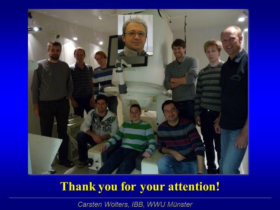
Thank you for your attention!
40 40
Ähnliche Präsentationen


 Natural Sources SNAP11.>")


 in Germany elective subject for medical students during.>")


